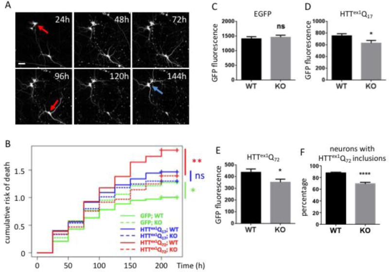
FIGURE 3.

Ube2W deficiency results in decreased Httex1Q72 inclusion formation and increased neuronal survival
A. Representative images from automated fluorescence microscopy. Primary cortical neurons from WT or Ube2W KO mice were transfected with mApple and Httex1Q72-EGFP, and survival was determined by repeated imaging at regular intervals. The last time at which the cell was noted to be alive (red arrows) was used as the time of death. Cells that survive the entire length of the experiment (blue arrow) were censored (analyzed as living cell at the end of experiment). Scale bar=25 μm.
B. Cumulative risk of death over time for WT and Ube2W KO neurons transfected with EGFP, Httex1Q17-EGFP and Httex1Q72-EGFP. Results were pooled from 16 wells per condition, with experiments performed in duplicate. ns, not significant; *, p<0.05; **, p<0.01. n>250.
C-F. Quantification of EGFP signals (C-E) or inclusion formation (F) from experiments as in panel A, 48 hours after transfection. Graphs show means +/− SEM; ns, not significant; *, p<0.05; ****, p<0.0001. n>232 for all analyses.
