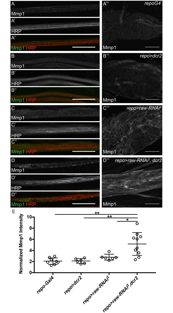
Fig 6. Mmp1 levels are increased upon raw knockdown.
(A-D'') Single slices of peripheral nerves of third instar larvae co-stained for Matrix metalloproteinase 1 (anti-Mmp1, green) and neurons (anti-HRP, red). All images acquired with 60x oil objective with same confocal settings. Scale bars are 25μm. (A-D) Mmp1 alone. (A'-D') HRP alone. (A''-D'') Merge of Mmp1 (green) and neurons HRP (red) (A‴-D‴) Single slices of the ventral nerve cord (VNC) of third instar larvae stained for Mmp1. All images acquired with 60x oil objective with same confocal settings. Scale bars are 50μm. (A-A‴) Control animals (repo-Gal4; n = 9). (B-B‴) Control animals overexpressing dcr2 (repo-Gal4> dcr2; n = 10). (C-C‴) Panglial raw knockdown (repo-Gal4>raw-RNAi2; n = 7). (D-D‴) Panglial raw knockdown in the presence of overexpressed dcr2 results in an altered Mmp1 levels in nerves and the VNC (repo-Gal4>raw-RNAi2, dcr2; n = 10). Posterior to the right. (E) Quantitation of average Mmp1 intensity in VNC relative to adjacent muscle tissue. **p<0.001 and *p<0.01 based on one-way ANOVA with post-hoc Tukey's test.

