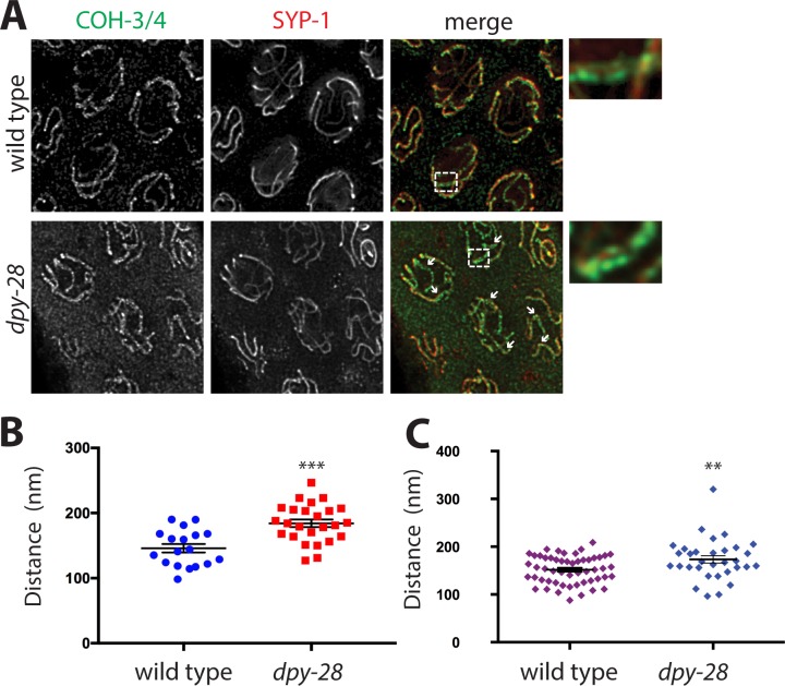
Fig 4. Defects in axial element separation condensin I mutants.
(A) STED microscopy images of cohesin (COH-3/4, green) and central element (SYP-1, red) localization on pachytene nuclei in wild type and dpy-28(tm3535) mutant males. Enlarged region is shown on the right. In both genotypes, two parallel tracks of cohesin along chromosomes can be resolved with a single SYP-1 track in between. Condensin I mutants show greater distances between the COH-3/4 tracks (arrows), indicating defects in chromosome axis formation. Because both COH-3/4 and SYP-1 signal intensities are reduced in dpy-28 mutants, we increased the laser power and detector gain to collect data, which resulted in higher levels of background in the mutants. (B) Distance measurements between COH-3/4 tracks in wild type (n = 18), and dpy-28 mutants (n = 25) at locations chosen randomly along chromosomes. (C) Distance measurements between COH-3/4 tracks in wild type (n = 52) and dpy-28 mutants (n = 32) at locations with a clear SYP-1 signal between the tracks. Asterisks indicate statistical significance by two-tailed, unpaired Student's t-test (*** indicates p<0.001, ** indicates p<0.01). Error bars indicate SEM.

