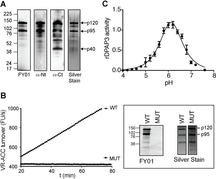
Fig 3. DPAP3 has proteolytic activity.
(A) Analysis of purified rDPAP3. Two main bands are detected by silver stain, both of which are strongly labelled by FY01 and recognized by the anti-Nt-DPAP3 and anti-Ct-DPAP3 antibodies. All other minor bands in the silver stain are also recognized by DPAP3 antibodies and represent degradation products that could not be separated during purification. (B) Measurement of VR-ACC turnover and FY01 labelling for WT and C504S MUT rDPAP3. Silver stain analysis shows equivalent amounts of protein were obtained from the purification of WT and MUT rDPAP3. (C) pH dependence of rDPAP3 activity measured at 10 μM VR-ACC (n = 3).

