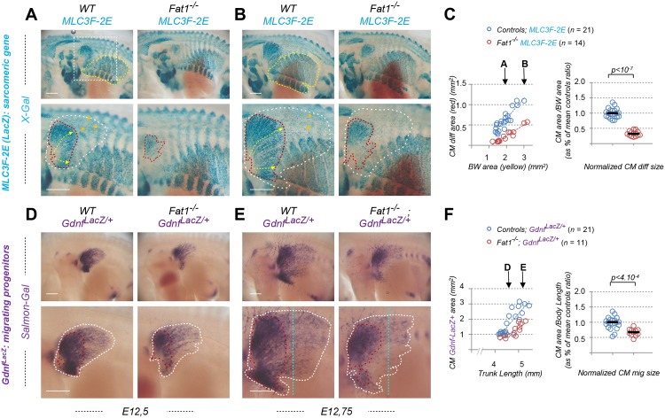
Fig 1. Fat1 knockout alters expansion of the subcutaneous muscle, CM.
Whole-mount β-galactosidase staining was performed using X-gal as substrate on embryos carrying the MLC3F-2E transgene (S1 Table) (A, B) or using Salmon-Gal as substrate on embryos carrying the GdnfLacZ/+ allele (S1 Table) (D, E). In each case, two successive stages are shown, E12.5 (A,D) and E12.75 (B, E), respectively with Fat1+/+ (left) and Fat1-/- (right) embryos, with the lower panels showing a higher magnification of the flank in which the CM muscle spreads. On upper panels in (A, B), the yellow dotted line highlights the BW muscles, the area of which is being measured. The white square highlights the area shown in lower panels. Lower panels: the red dotted line highlights the area covered by differentiating MLC3F-2E+ muscle fibers constituting the CM muscle in (A, B), also matching an area of higher GdnfLacZ intensity in (D, E); the white dotted lines highlight the area corresponding to the full shape of the GdnfLacZ+ area in (D, E), in which a low density of blue (MLC3F-2E+) nuclei can also be observed in (A, B). (C) Quantifications of the relative expansion of the MLC3F-2E+ CM differentiated area. Left plot: for each embryo side, the area of differentiated CM is plotted relative to the BW area. Arrows represent the stages shown in (A) and (B), respectively. Right plot: for each embryo, the CM area/BW area was normalized to the median ratio of control embryos. Blue dots: Fat1+/+; MLC3F-2E (n = 21); red dots: Fat1-/-; MLC3F-2E (n = 14). Underlying data are provided in S1 Data. (F) Quantifications of the relative expansion of the GdnfLacZ+ area. Left plot: for each embryo side, the GdnfLacZ+ area is plotted relative to the length of the trunk (measured between two fixed points). Arrows represent the stages shown in (D) and (E), respectively. Right plot: for each embryo, the CM area/trunk length was normalized to the median ratio of control embryos. Blue dots: Fat1+/+; GdnfLacZ/+ (n = 21); red dots: Fat1-/-; GdnfLacZ/+ (n = 11). Underlying data are provided in S1 Data. Scale bars: 500 μm. BW, body wall; CM, cutaneous maximus; Gdnf, glial cell line-derived neurotrophic factor; MLC3F-2E, Mlc3f-nLacZ-2E line (S1 Table); Salmon-Gal, 6-Chloro-3-indolyl-β-D-galactopyranoside, substrate for β-galactosidase activity; WT, wild-type; X-gal, 5-bromo-4-chloro-3-indolyl-β-D-galactopyranoside, substrate for β-galactosidase activity.

