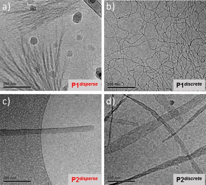
Figure 3.

CryoTEM images at 25000 magnification of (a) P1disperse and (b) P1discrete, both at 5 mg mL–1 in water with 10% THF, and (c) P2disperse and (d) P2discrete both at 2 mg mL–1 in water with 10% THF. Large dark round particles in “a” are crystalline ice particles and not part of the sample. The images were recorded at 10 μm (a, b, d) and 5 μm defocus (c). For corresponding low-magnification images see Figure S17.
