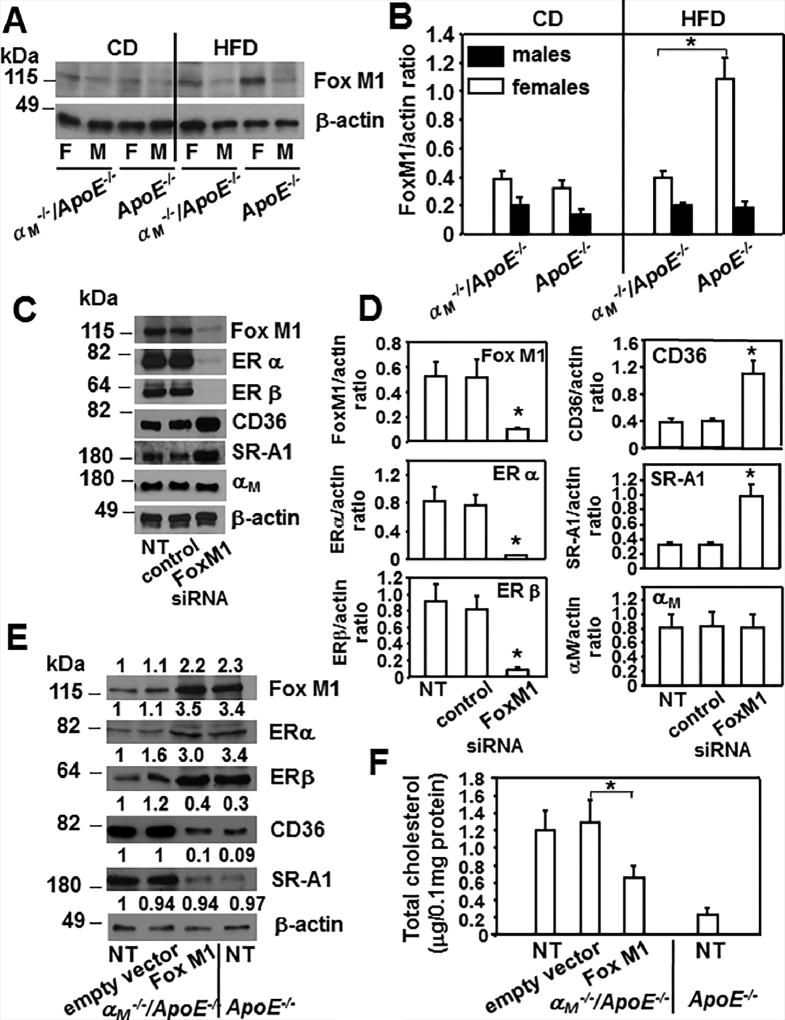
Figure 8. Fox M1 regulates expression of ERs in macrophages and it is reduced exclusively in female αM−/−/ApoE−/− macrophages.
(A) Western blot analysis of peritoneal macrophages derived from αM−/−/ApoE−/− and ApoE−/− mice fed CD or HFD probed with Ab to FoxM1 and β-actin. Images are representative of 3 independent experiments (B) Densitometric quantification of Western blots from A. (*P<0.05, female αM−/−/ApoE−/− vs female ApoE−/− mice, n=6). (C) Western blot analysis of macrophages derived from female ApoE−/− mice that were untreated or treated with FoxM1 siRNA or control siRNA. Western blots were probed with indicated Abs and β-actin as loading control. (D) Densitometric quantification of Western blots from C. (*P<0.05, FoxM1 siRNA treated macrophages vs untreated or control-siRNA treated cells, n=6). Three independent experiments were performed. (E) Western blot analysis of female αM−/−/ApoE−/− macrophages that were untreated or transfected with FoxM1 pcDNA3.1 or empty vector pcDNA3.1 as described in Materials and Methods. Also lysates of untreated female ApoE−/− macrophages were included as control. Western blots were probed with indicated antibodies and anti-β-actin for loading control. Densitometric quantification and values of band densities are shown above each blot and they are calculated relative to the control sample of untreated αM−/−/ApoE−/−, which has been assigned value 1. (F) Foam cell formation was measured as described in Materials and Methods in macrophages treated as in E. (*P<0.05, FoxM1 transfected αM−/−/ApoE−/− macrophages vs transfected with empty vector, n=8). Three independent experiments were performed.

