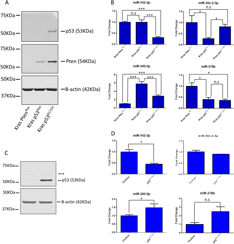
Fig. 2. Validation of microRNA expression in primary cell lines and Kras p53flox cells with ectopic expression of mutant p53R172H.
a Primary cell lines with the same genotype as the primary tissue samples used in Fig. 1 were analysed for protein expression by western blot. b The expression of the microRNAs identified as being dysregulated in p53R172H-expressing tissues was investigated in these cell lines using the ΔΔCT method with U6 snRNA as the reference gene. The results represent the mean of three biological repeats; the error bars represent the maximum relative quantity. A two-sample, two-tailed, paired t-test was used to compare the ΔCT values from each group. Statistical significance is represented as *p < 0.05 and ***p < 0.005. c Kras p53flox with ectopic expression of p53R175H and a control cell line stably expressing an empty vector were analysed by western blot. The * represents a non-specific band of unknown origin. d Expression of the microRNAs identified as being dysregulated in the Kras p53R172H tissues was interrogated by RT-qPCR in these cell lines using the ΔΔCT method with U6 snRNA as the reference gene. The results represent the mean of three biological repeats with the error bars representing the maximum relative quantity. A two-sample, two-tailed, paired t-test was used to compare the ΔCT values from each group. Statistical significance is represented as *p < 0.05

