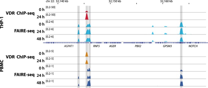
Figure 2.
Vitamin D receptor (VDR) binding and chromatin opening of the 1-acylglycerol-3-phosphate O-acyltransferase 1 (AGPAT1) locus in vitro and in vivo. Top: THP-1 cells were stimulated for 0, 24, and 48 h with 1,25(OH)2D3 and VDR chromatin immunoprecipitation sequencing (ChIP-seq) and formaldehyde-assisted isolation of regulatory elements sequencing (FAIRE-seq) were performed. Bottom: in an analogous in vivo experiment (phase II context of the VitDbol study) one individual was challenged with a vitamin D3 bolus (2,000 µg). The average raise in 25(OH)D3 serum concentrations at days 1 and 2 after the vitamin D3 bolus was 11.9 and 19.4 nM, respectively. Peripheral blood mononuclear cells (PBMCs) were isolated before (day 0) and at days 1 (24 h) and 2 (48 h) and VDR ChIP-seq and FAIRE-seq were performed. The integrative genomics viewer browser was used to visualize the AGPAT1 gene locus. The peak tracks represent mergers of each three biological repeats. Gene structures are shown in blue.

