FIGURE 4.

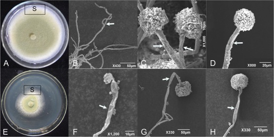
Scanning electron micrographs showing the healthy (A–D) and shriveled conidiophores (E–H) of A. flavus on PDA amended with culture filtrates of S. yanglinensis 3-10 at 2% (v/v). S, sampling site. The arrows mean hyphae or conidiophore.

Scanning electron micrographs showing the healthy (A–D) and shriveled conidiophores (E–H) of A. flavus on PDA amended with culture filtrates of S. yanglinensis 3-10 at 2% (v/v). S, sampling site. The arrows mean hyphae or conidiophore.