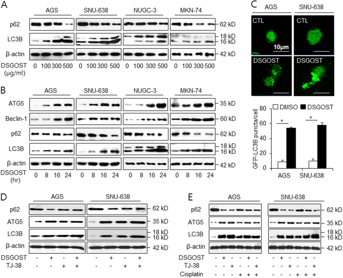
Fig. 2. DSGOST activates autophagy in gastric cancer cell lines.
a, b AGS and SNU-638 were treated with DSGOST in a dose- (100, 300, and 500 μg/mL; 24 h) and a time-dependent manner (8, 16, and 24 h; 500 μg/mL), and the control cells were treated with DMSO. Sampling of total lysates was conducted by western blot assay to identify the activation of autophagy markers in a DSGOST dose- and time-dependent manner. c AGS and SNU-638 cells transfected by the pEGFP-LC3 vector were treated with DMSO or DSGOST (500 μg/mL) for 8 h. Fluorescence microscopy analysis calculated by puncta of LC3B staining. The graph indicates the number of cells with GFP-LC3B puncta; *p < 0.05. d Western blot assay of LC3B, ATG5, and p62 level in DSGOST (500 μg/mL, 24 h) and/or TJ-38 (500 μg/mL, 24 h)-treated AGS and SNU-638 cells. e After AGS and SNU-638 cells were treated with DSGOST (500 μg/mL) or TJ-38 (500 μg/mL) in combination with cisplatin (5 µM) for 24 h, protein samples were loaded to perform western blotting for LC3B, ATG5, and p62. β-actin was used as the protein loading control

