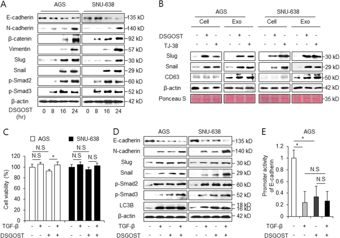
Fig. 6. DSGOST induces EMT in gastric cancer.
a AGS and SNU-638 cells were treated with DSGOST (500 μg/mL) in a time-dependent manner (0, 8, 16, and 24 h). After DSGOST treatment, extracted total proteins were subjected to western blotting using antibodies for EMT markers, such as E-cadherin, N-cadherin, β-catenin, vimentin, Slug, Snail, and Smad signaling, including p-Smad2 and p-Smad3. b AGS and SNU-638 cells were treated with DSGOST or TJ-38 and exosomes were collected from cell supernatants. Exosomes and cell lysates were quantified by Ponceau S staining. Exosomes were also examined by western blotting using the exosome marker CD63 and EMT markers, such as Snail and Slug. c, d Cell viability and western blotting analysis of EMT markers and Smad signaling in AGS and SNU-638 cells after TGF-β1 (5 ng/mL, 24 h) and DSGOST (500 μg/mL, 24 h) treatment; *p < 0.05. e AGS cells were transfected with a reporter luciferase vector containing an E-cadherin promoter (−368~+51), and were treated with TGF-β1 (5 ng/mL, 24 h) or/and DSGOST (500 μg/mL, 24 h). The luciferase activity was normalized to the activity of Renilla luciferase; *p < 0.05

