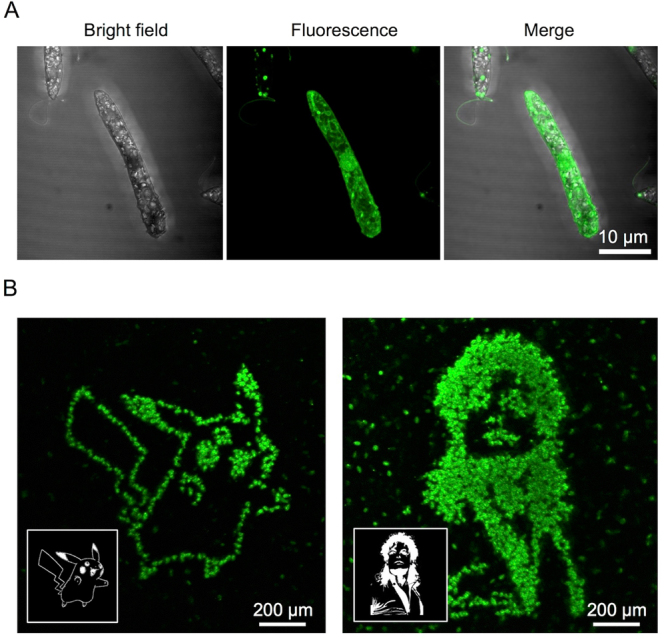
Figure 4.

Femtosecond laser photoporation of E. gracilis cells at single-cell resolution. (A) Bright-field and fluorescence images of an E. gracilis cell 20 min after the photoporation with the FPBP. (B) Fluorescence images of E. gracilis cells 20 min after the spatially patterned photoporation with the same aptamer. The patterned photoporation was performed on the cells in the black and white patterns of Pikachu (left) and Michael Jackson (right) as shown in the insets. Each fluorescent dot corresponds to a single E. gracilis cell into which the aptamer was injected and bound to intracellular paramylon. The images firmly show the demonstration of the photoporation with the single-cell resolution.
