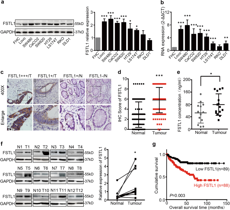
Fig. 1. FSTL1 is up-regulated in CRC and correlates with poor prognosis.
Expression analyses of FSTL1 protein and mRNA in CRC cell lines by a western blotting and b qPCR. FSTL1 protein levels were normalised to the relative expression of FHC. FSTL1 mRNA expression was quantified by qPCR and normalised to GAPDH. Error bars represent the mean ± S.D. (n = 3). c Immunohistochemistry (IHC) staining in 130 paraffin-embedded CRC tissues sections. (i) Strong expression (+++) in CRC. (ii) Weak expression (+) in CRC. (iii) Weak expression (+) in adjacent normal tissues. (iv) Scored negative for expression (−) in adjacent normal colorectal tissues. d Comparison of FSTL1 expression scores in CRC tissues (Tumour) with adjacent non-tumour tissues (Normal), P < 0.0001. e Elisa assay of FSTL1 in serum from healthy donors (Normal, n = 13) and CRC patients (Tumour, n = 15), P = 0.046. f Western blotting analysis of FSTL1 in 12 CRC tissues (T) and paired normal colorectal mucosa (N). Quantification of protein expression shown in right was normalised to GAPDH, P = 0.0178. g Kaplan–Meier survival analysis of GEO Database (GSE17536, n = 177) according to the expression of FSTL1, P = 0.003 (Log-rank test). *P < 0.05, **P < 0.01, ***P < 0.001

