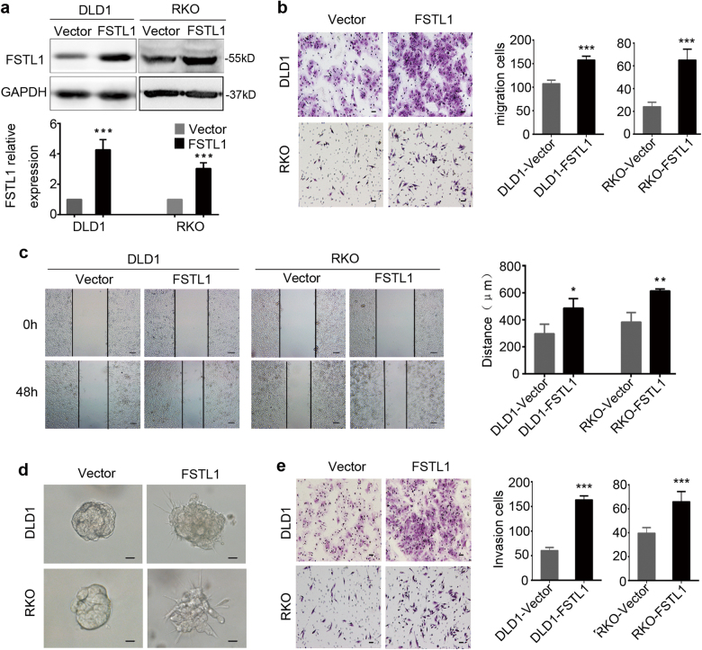
Fig. 2. FSTL1 promotes CRC cells migration and invasion in vitro.
a DLD1 and RKO cells were stably transfected with vector (Vector) or FSTL1 lentivirus (FSTL1). Western blotting analysis was performed to detect the expression of FSTL1 (upper panel). FSTL1 protein levels were normalised to the relative expression of Vector group respectively, P < 0.001 (lower panel). Error bars represent the mean ± S.D. (n = 3). b The migratory cells of trans-well assay were counted under microscope in five randomly selected fields (scale = 50 μm). Representative photographs (left) and quantification (right) are shown, both P < 0.0001. Error bars represent the mean ± S.D. (n = 5). c Wound healing assay was performed to evaluate the motility in DLD1 cells, P = 0.03 and RKO cells, P = 0.0052 (scale = 100 μm). Error bars represent the mean ± S.D. (n = 3). d Three-dimensional culture assays was performed to observe the effect of FSTL1 on cell invasion (scale = 20 μm). FSTL1 overexpressing cells were extended protuberances to the Matrigel matrix, and the control cells formed tight spherical colonies. e The invasive cells of Matrigel-coated Boyden chamber invasion assay were counted under microscope in five randomly selected fields (scale = 50 μm). Representative photographs (left) and quantification (right) are shown, both P < 0.0001. Error bars represent the mean ± S.D. (n = 5). *P < 0.05, **P < 0.01, ***P < 0.001

