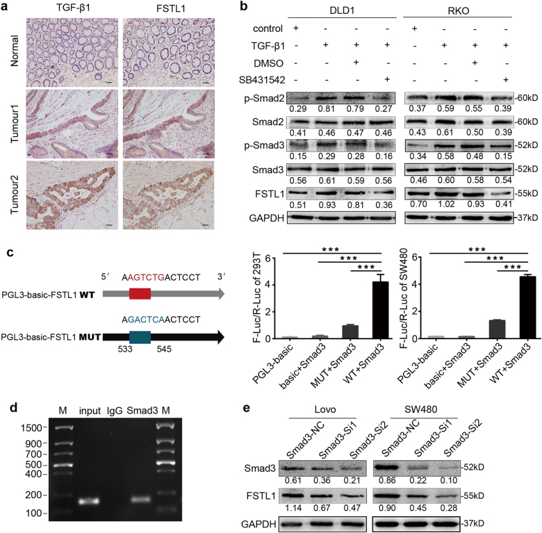
Fig. 5. TGFβ1-Smad2/3 signalling pathway regulates the expression of FSTL1 through activating the transcriptional activity of Smad3 in human CRC.
a Representative TGF-β1 and FSTL1 immunohistochemistry staining photographs of normal tissue (Normal) and tumour tissue samples (Tumour 1, Tumour 2), (×200, scale = 50 μm). The TGF-β1 and FSTL1 were spatially correlated. b TGF-β1 activated Smad2/3 signalling and enhanced FSTL1 expression in DLD1 and RKO cells by western blotting. SB431542 attenuated the expression of P-smad2, P-smad3 and FSTL1. c Schematic of the FSTL1 promoter luciferase construct is depicted with the locations of binding site and the sequences of mutation (left). Luciferase activities in HEK293T and SW480 cells were examined after transfecting with Smad3 (right), each P < 0.001. Error bars represent the mean ± S.D. of the ratio of firefly and renilla luciferase signals (n = 3). d Smad3 binding on the promoter region of FSTL1 was assessed by ChIP assay. Immunoprecipitation from SW480 cells using Smad3 antibody or rabbit immunoglobulin G (IgG). PCR from the IP samples using FSTL1-specific primers. e Western blotting analysis of Smad3 and FSTL1 in Lovo and SW480 cells, after using smad3-siRNA mediated RNA interference. *P < 0.05, **P < 0.01, ***P < 0.001

