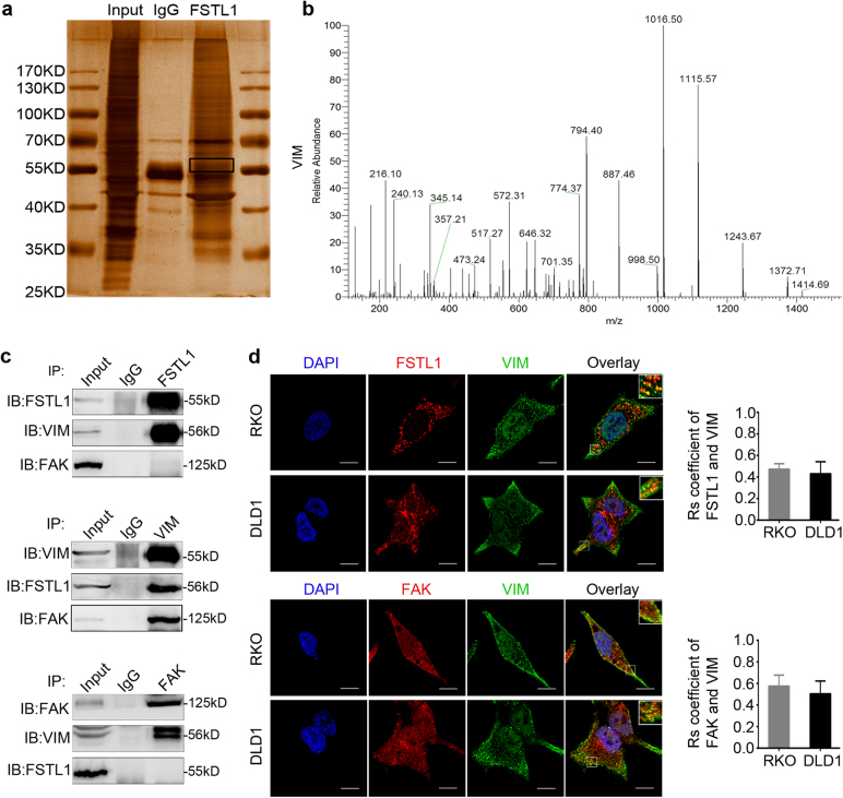
Fig. 7. VIM is an interactive factor of FSTL1.
a Immunoprecipitation and silver staining was performed by using the whole proteins SW480 cells with anti-FSTL1 antibody. b The spectrogram of differential protein band is identified as VIM (Quadrupole Mass Spectrometer). c Coimmunoprecipitations were performed to validate the interaction among FSTL1, VIM, and FAK in SW480 cells. d FSTL1 (red, upper panel) or FAK (red, lower panel) co-localizate with VIM (green) in RKO and DLD1 cells was assessed by laser-scanning confocal microscopy, respectively (×1800, scale = 10 μm). DAPI (blue) stains nuclei. The image in white pane of the upper right corner of overlay is a partial enlarged detail. Error bars in the histogram represent the mean ± S.D. of the colocalisation coefficients of VIM and FSTL1 (upper panel) or the colocalisation coefficients of VIM and FAK (lower panel) in RKO cells and DLD1 cells (n = 10)

