Figure 2.
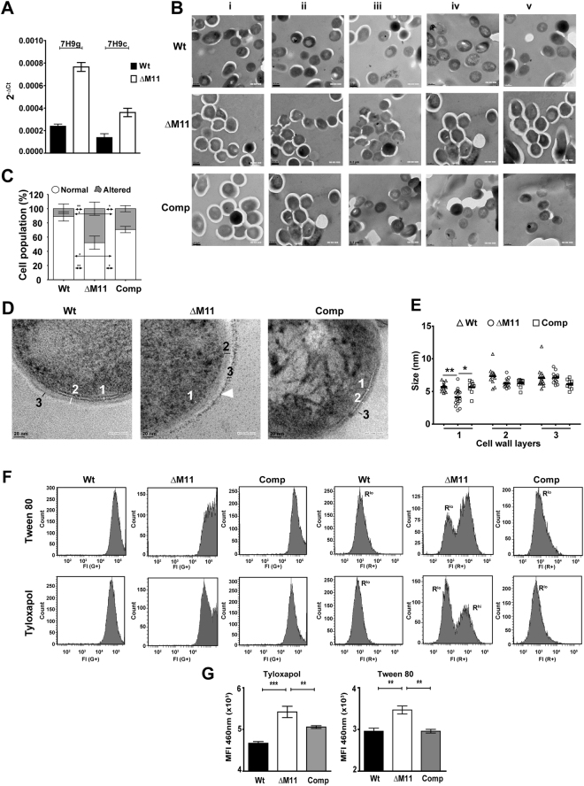
Loss of mmpL11 in Mtb results in changes of the gross cell wall ultrastructure and in membrane integrity in Tyloxapol: (A) Expression of sigE gene in the Wt and mutant strain by qRT PCR in media containing 0.05% Tyloxapol - 7H9g (7H9 containing 0.5% glycerol) and 7H9c (0.5% glycerol with ADC). The fold change in Ct values with respect to 16 s rRNA is depicted. (B,D) Representative TEM micrographs (5 different fields are shown as i-v) of Wt, ΔM11 and Comp strains grown in media containing 0.05% Tyloxapol. Scale bars of 0.2 µm (B) and 20 nm (D) are shown. (C) Quantitation of cells in the samples with intact morphology (round) and rhomboid morphology (outlines) from 3 independent experiments. (E) Thickness of cell wall layers of Wt, ΔM11 and Comp Mtb strains. Each symbol represents the micrograph of one cell, median values are depicted. (F) Analysis of membrane polarity in media containing either 0.05% Tween 80 or Tyloxapol by DiOC2(3) staining and FACS. Fluorescence intensities (FI) at 497 nm (G+) and 615 nm (R+) are represented as histograms. Rlo and Rhi represent the tow populations with low and high red intensities, respectively. (G) Uptake of Hoechst-33342 by the Mtb strains. Data represents mean fluorescence intensities (MFI) ± SD values of triplicate wells in a representative experiment of n = 3.

