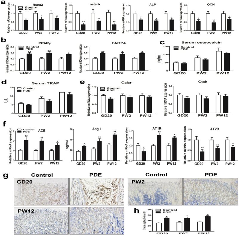
Fig. 3. Effects of prenatal dexamethasone exposure (PDE) on osteogenic differentiation and local renin–angiotensin system (RAS) in F1 male offspring.
a RT-qPCR analysis of gene expression of osteogenic differentiation markers, including Runx2, osterix, alkaline phosphatase (ALP), and osteocalcin (OCN) in bone tissue from gestational day (GD) 20 to postnatal week (PW) 12. b RT-qPCR analysis of gene expression of adipogenic differentiation markers, including peroxisome proliferator-activated receptor gamma (PPARγ) and fatty acid-binding protein 4 (FABP4) in bone tissue from GD20 to PW12. c ELISA analysis of serum osteocalcin from GD20 to PW12. d Analysis of serum TRAP activity from GD20 to PW12. e RT-qPCR analysis of gene expression of osteoclast differentiation markers in bone tissue from GD20 to PW12. f RT-qPCR analysis of gene expression of RAS, including angiotensin-converting enzyme (ACE), angiotensin receptors (ATRs), and ELISA analysis of angiotensin II (Ang II) production in bone tissue from GD20 to PW12. g Representative immunostaining images of ACE in bone tissue from GD20 to PW12. h Quantitative immunostaining analysis of the mean optical density of ACE from GD20 to PW12. Mean ± S.E.M., n = 8 per group, *P < 0.05, **P < 0.01 compared with the control

