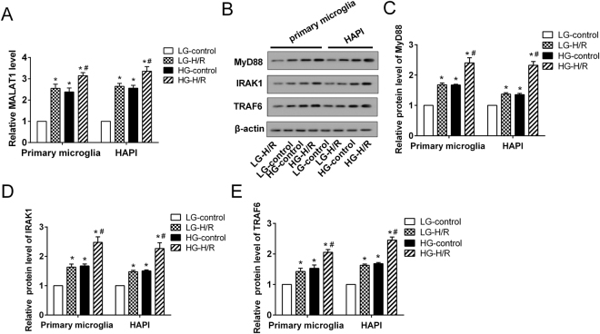
Figure 3.
The expression of MALAT1, MyD88, IRAK1 and TRAF6 in an in vitro model of DM-I/R. The rat primary microglia and microglia line HAPI were cultured to establish the cell model of DM-I/R with high glucose (HG) and hypoxia-reoxygenation (H/R). The cells were divided into 4 groups: LG-control (low glucose treatment, 5.5 mM), LG-H/R (LG and H/R treatment), HG-control (HG treatment, 30 mM), HG-H/R (HG and H/R treatment). (A) The expression of MALAT1. (B) Representative Western blot analysis of MyD88, IRAK1, TRAF6 protein. The relative expressions of (C) MyD88, (D) IRAK1, (E) TRAF6 protein were measured. *P < 0.01 vs. LG-control; #P < 0.01 vs. LG-H/R.

