Figure 4.
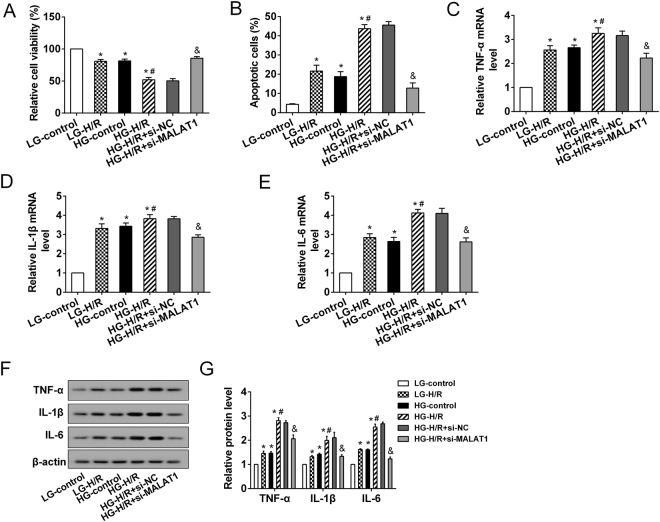
The effects of MALAT1 on microglia in vitro. The HAPI cells were divided into 6 groups: LG-control, LG-H/R, HG-control, HG-H/R, HG-H/R + si-NC, HG-H/R + si-MALAT1. (A) The relative cell viability was detected with MTT assays. (B) The apoptotic cell was assessed by flow cytometry. (C–E) The expression of proinflammatory cytokines (TNF-α, IL-1β and IL-6) was measured at mRNA levels. (F) Representative Western blot analysis of TNF-α, IL-1β and IL-6. (G) The relative expressions of TNF-α, IL-1β, and IL-6 protein were measured. *P < 0.01 vs. LG-control; #P < 0.01 vs. LG-H/R; &P < 0.01vs. HG-H/R + si-NC.

