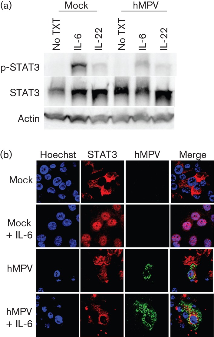
Fig. 4.

Inhibition of STAT3 phosphorylation and translocation in hMPV-infected NHBE cells. NHBE cells were infected at an m.o.i. of 0.5. At 48 h p.i., cells were treated with 20 ng IL-6 ml −1 for 30 min. (a) Cell lysates were analysed by Western blot analysis with antibodies against p-STAT3, STAT3 or actin. (b) Cells were fixed with 4 % paraformaldehyde and stained for viral antigen (green) and STAT3 (red). Nuclei were counterstained with Hoechst (blue). Results are representative of two independent experiments. No TXT, no cytokine (IL-6 or IL-22) treatment.
