Figure 7.
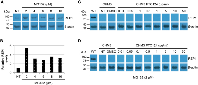
Effect of MG132 treatment on wild-type and CHM3 iPSC-derived RPE. (A) Western blot analysis of REP1 expression following treatment of wild-type iPSC-derived RPE with increasing concentrations of MG132. β-actin serves as a loading control. Bands were cropped from the same gel with different exposure times for REP1 and β-actin. (B) Quantification of REP1 expression in panel A shows an increase at all concentrations used with the highest levels observed at 2 µM (~5-fold higher than non-treated (NT) cells). Western blot analysis of REP1 expression in non-treated (NT), DMSO-treated or PTC124-treated CHM3 iPSC-derived RPE in the absence (C) or presence (D) of MG132. β-actin serves as a loading control. No expression could be seen in CHM3 iPSC-derived RPE as compared to wild-type (WT) RPE regardless of the treatment used. In panels C and D, bands were cropped from the same gel with different exposure times for REP1 and β-actin.

