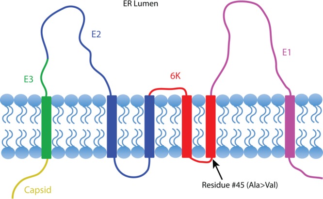
FIG 2.

Threading of EEEV structural polyprotein through an endoplasmic reticulum membrane. Structure proteins are colored separately: Capsid in yellow, E3 in green, E2 in blue, 6K in red, and E1 in magenta. The positive selection pressure site, residue 45 in 6K, is indicated by a black arrow.
