FIG 1.
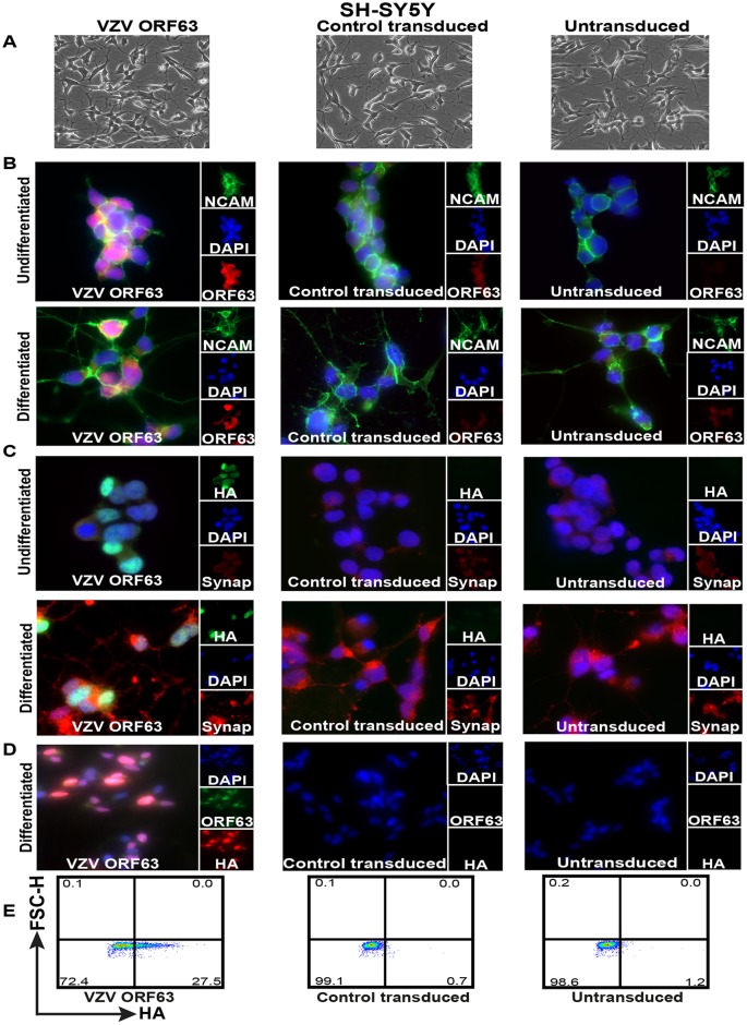
Validation of HA-tagged VZV ORF63-expressing differentiated SH-SY5Y cells. (A) SH-SY5Y cells (1.5 × 10⁶) were transduced with VZV ORF63 or CT pseudoviruses and selected with 0.4 mg/ml G418 for 10 days to create VZV ORF63, CT, and untransduced SH-SY5Y cells. These cells were differentiated using 10 μM ATRA for 5 days and 50 ng/ml BDNF for 4 days. The cells were imaged by light microscopy at the end of the differentiation protocol; the images are shown at ×20 magnification. (B to D) VZV ORF63, control transduced, and untransduced SH-SY5Y cells (1 × 10⁵) were differentiated on Matrigel-coated coverslips (13 mm; Knittel glass), fixed with 4% PFA, and stained for VZV ORF63 and NCAM (B), HA and synaptophysin (C), or VZV ORF63 and HA (D). The cells were counterstained with nuclear DAPI (blue) and were visualized by fluorescence microscopy. The images are shown at ×20 (D) or ×63 (B and C) magnification. (E) Additionally, 5 × 10⁵ differentiated VZV ORF63, CT, and UT SH-SY5Y cells were fixed, permeabilized, stained for HA, and analyzed via flow cytometry. All the data presented are representative of three biological replicates, except for IFA staining (B and C), which is representative of two biological replicates.

