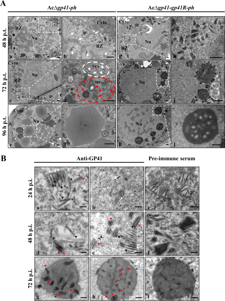
FIG 3.
EM analysis of ODV morphogenesis and GP41 localization. (A) EM analysis of virion morphogenesis in recombinant virus-transfected cells. Sf9 cells were transfected with AcΔgp41-ph (a to f) or AcΔgp41-gp41R-ph (g to l) and fixed at 48, 72, and 96 h p.t. Ultrathin sections of the cells were observed by TEM. For each panel on the left, the right picture show an enlarged view of the boxed region. White arrows and red arrows indicate nucleocapsids and MVs, respectively. Nu, nucleus; Cyto, cytoplasm; NCs, nucleocapsids. Bars, 500 nm. (B) IEM analysis of GP41 localization in infected cells and virions. Sf9 cells were infected with control virus at an MOI of 5 TCID50 units/cell and harvested at 24, 48, and 72 h p.i. The cells were probed with anti-GP41 pAb as the primary antibody and goat anti-rabbit IgG coated with gold particles (10 nm) as the secondary antibody. Cell sections were also detected with preimmune rabbit serum as a control group. The samples were observed by TEM. Arrowheads indicate the location of GP41. Bars, 200 nm.

