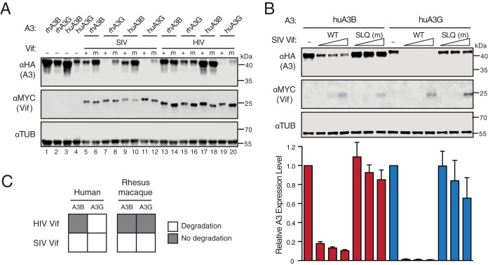
FIG 1.
SIVmac239 Vif degrades both human APOBEC3B and APOBEC3G. (A) Immunoblots of 293T cells expressing the indicated rhesus macaque (rh) or human (hu) A3 enzymes together with SIVmac239 Vif, HIV-1IIIB Vif, or SLQ-AAA mutant (m) derivatives. A3 enzymes were detected using an anti-HA antibody for a C-terminal HA epitope tag. Vif was detected using an anti-MYC antibody for a C-terminal MYC epitope tag, and anti-α-tubulin was used as a loading control. (B) Bar graphs for quantification (bottom) of immunoblots (top and not shown) of 293T cells expressing fixed amounts of the indicated human A3 enzymes together with an empty vector control (−) or various amounts of SIVmac239 Vif (WT) or a SLQ-AAA mutant (m) derivative (analyzed with the same antibodies as for panel A). A3 expression levels were determined by quantifying band intensities normalized to those of the corresponding Vif-null condition. Each error bar represents the standard deviation for three biological replicates. (C) Schematic summarizing the immunoblotting results from panels A and B. Open squares represent a functional Vif-A3 interaction evidenced by A3 degradation.

