. 2018 May 28;19(3):406–415. doi: 10.4142/jvs.2018.19.3.406

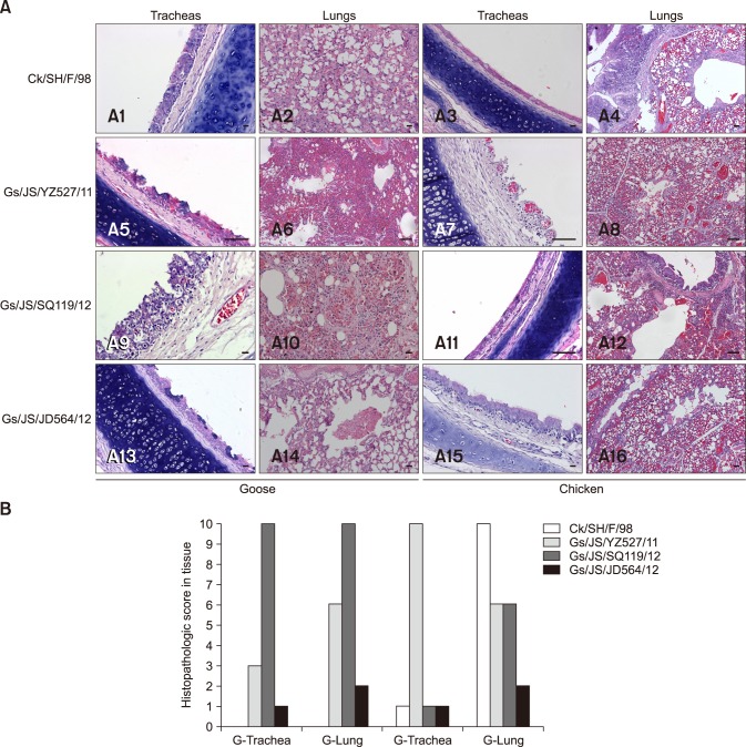
Fig. 3. (A) Histopathology of SPF chickens and Ck/SH/F/98, Gs/JS/YZ527/11, Gs/JS/SQ119/12, and Gs/JS/JD564/12 infected geese. Lungs and tracheas were collected at 4 days post inoculation fixed in 10% formalin, embedded in paraffin, and sectioned. (B) Pathological changes in trachea and lung in chickens at 4 days after inoculation with the isolated viruses. Tissue sections were observed, and histopathological changes were scored as follows. For trachea, 0, normal; 1, congestion; 2, cilia loss; 3, a few inflammatory cell infiltration; 7, a lot of inflammatory cell infiltration. For lung, 0, normal; 1, congestion; 2, hemorrhage; 3, inflammatory cell infiltration in bronchial submucosa; 7, a lot of inflammatory cell infiltration in bronchial submucosa and alveolus. Average values for three birds are shown. Data are representative of three independent experiments. G, goose; C, chicken. Scale bars = 5 µm (A9 and A10), 10 µm (A1, A3, and A15), 20 µm (A2, A14, and A16), 50 µm (A4–8, A11–13).

Fig. 3