
Fig. 3. Effect of OMT on apoptotic proteins in L02 cells.
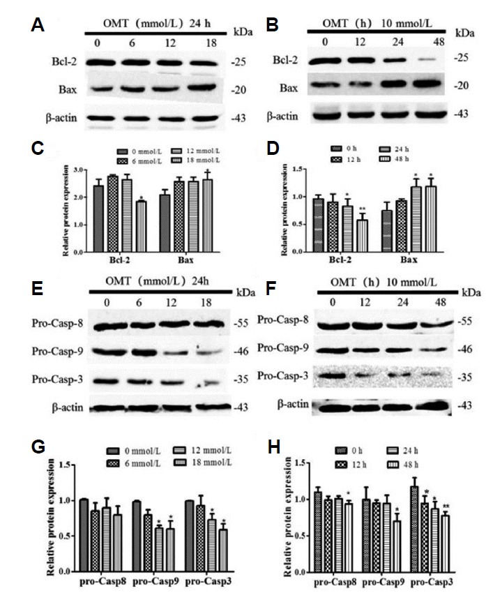
Cells were cultured with 0, 6, 12, 18 mmol/L of OMT for 24 h or 10 mmol/L OMT at 12, 24 and 48 h, and then all proteins were isolated from the cells. The expression of Bcl-2, Bax (A, B) and pro-caspase-9/-8/-3 (E, F) proteins were detected by western blotting and normalized to β-actin. Relative band intensities were used in order to quantify protein expression levels (C, D, G, and H). *P < 0.05, **P < 0.01 vs the control group.