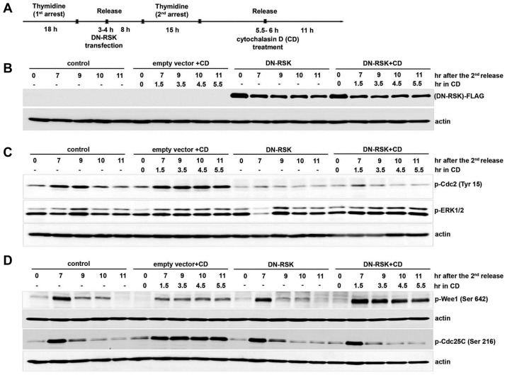
Fig. 3. Overexpression of dominant-negative RSK partially releases the cell cycle delay by actin dysfunction.
(A) The layout shows how the experiment was performed. IMR-90 cells were synchronized with 2 mM thymidine and transfected with DN-RSK or empty vector as described in the Materials and Methods. Cells were treated with 5 μM CD or only DMSO as a control at 5.5–6 h after the second release and collected at each indicated time point after the second release. Each cell lysate was resolved by 8% SDS-PAGE and blotted with (B) anti-FLAG and anti-actin, (C) p-ERK1/2 and p-Cdc2 (Tyr 15), (D) p-Cdc25C (Ser 216), and p-Wee1 (Ser 642). Each membrane was reprobed with anti-actin as a loading control.

