Figure 2.

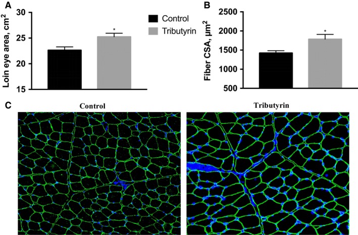
At 58 days of age, cross‐section of Longissimus dorsi (LD) muscle was taken at the 12th rib and utilized for immunohistochemical analysis to determine fiber cross‐sectional area (FCA). Values depicted are based off pooled neonatal control (C, n = 12) or tributyrin (T, n = 12) treatment groups. (A) The cross‐sectional area of the LD at the 12th rib (loin eye). (B) LD muscle FCA as determined by immunohistochemistry. (C) Immunohistochemical analysis of FCA of the LD muscle. Muscle fibers were cryosectioned and stained with anti‐dystrophin to visualize sarcolemma (green), >400 fibers/animal were counted using Zeiss ZEN Pro automated image analysis. Nuclei were visualized with DAPI. Significance was declared at P < 0.05 (*).
