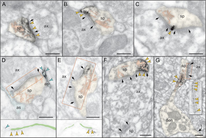
Figure 3.
Postsynaptic expression of mGluR3 in monkey dlPFC. mGluR3 is prominently expressed in dendritic spines, both within the synaptic active zone (A) and perisynaptically (B,C); label in C is found at the central perforation of a perforated synapse. (D,E) The framed images are edited to facilitate receptor visualization at the synapse (the synaptic cleft is marked in green). Note in D the typical mGluR3 localization in PAPs. (F,G) mGluR3 is additionally expressed at nonsynaptic spine membranes. In F, one section of a perforated synapse is additionally labeled. Extrasynaptic mGluR3 in G is found next to the spine apparatus (pink-pseudocolored) in the spine neck of a prototypical thin spine; a second spine, not shown in its entirety, emanates from the parent dendrite (curved arrow). The enlarged frame in G shows the common postsynaptic expression of mGluR3 at the synapse. Labeled spines (sp) and astrocytes (as) are pseudocolored for clarity; color-coded arrowheads point to mGluR3; synapses are between arrows. ax, axon; den, dendrite. Scale bars, 200 nm.

