Figure 1.
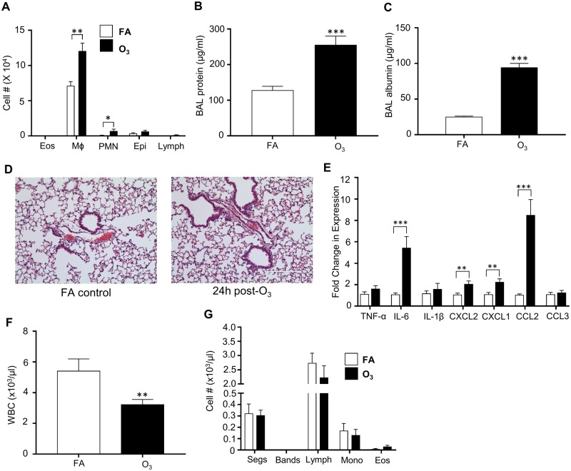
O3 exposure increases pulmonary inflammation and injury while decreasing circulating immune cells. Inflammatory response to O3 exposure in lung tissue. Mice were exposed to 1 ppm O3 or filtered air (FA) for 3 h and necropsied 24 h following exposure. Bronchoalveolar lavage (BAL) was analyzed for A, cell differentials (n = 10 per group). B, total protein (n = 10 per group), and C, increases in BAL albumin concentrations (n = 3–5 per group). Lung tissue was stained with D, hematoxylin and eosin to examine pulmonary damage. E, Whole lung homogenate was analyzed for gene expression of cyto/chemokines which was normalized to 18S (n = 5 per group). Mice were also analyzed for F, total white blood cell (WBC) counts (n = 10 per group) and G, cell differentials (n = 5 per group). *p < .05; **p < .01; ***p < .001.

