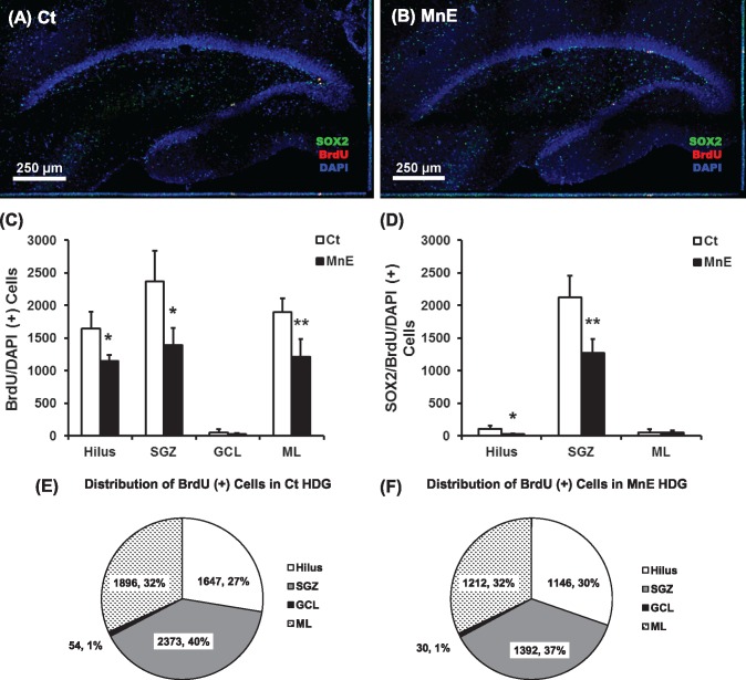
Figure 2.
Cell proliferation in adult hippocampal dentate gyrus (HDG) with or without subchronic Mn exposure. A, Adult rats received saline as the control (Ct). B, Rats received Mn injections as the Mn-exposed group (MnE). See Figure 1A for detailed experimental design. Newborn cells were labeled with BrdU (red). 4′,6-Diamidino-2-phenylindole (DAPI) (blue) was used to stain nuclei and delineate the structure of the HDG. C, Quantification of BrdU/DAPI-labeled cells in the HDG subregions (ie, hilus, subgranular zone [SGZ], granule cell layer [GCL], and molecular layer [ML]) of both hemispheres following in vivo subchronic Mn exposure. D, Quantification of Sox2/BrdU/DAPI(+) cells in the HDG of both hemispheres following subchronic Mn exposure. Data represent mean ± SD, n = 4. *p < .05, as compared with controls. E, Percentage distribution of BrdU-labeled in the control HDG subregions. F, Percentage distribution of BrdU-labeled in the Mn-exposed HDG subregions.

