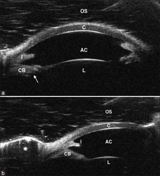
Figure 2.

(a) UBM of the OS showing the cornea (C), deep AC, iris stump (I), enlarged CB with anterior rotation of its posterior part, the ciliary processes (arrow) and the lens (L). (b) UBM of the OS showing the cornea (C), deep AC, iris stump (I), enlarged CB with anterior rotation of its posterior part, the lens (L), and the formed bleb (asterisk). UBM: Ultrasound biomicroscopy, OS: Left eye, AC: Anterior chamber, CB: Ciliary body
