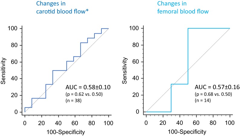
Fig. 3.
Receiver operating characteristic curves describing the ability of changes in carotid femoral blood flows to detect a positive response of cardiac index to a passive leg raising test (increase ≥ 10%). AUC area under the curve. Asterisks results are provided for carotid blood flow measured by the velocity time integral method

