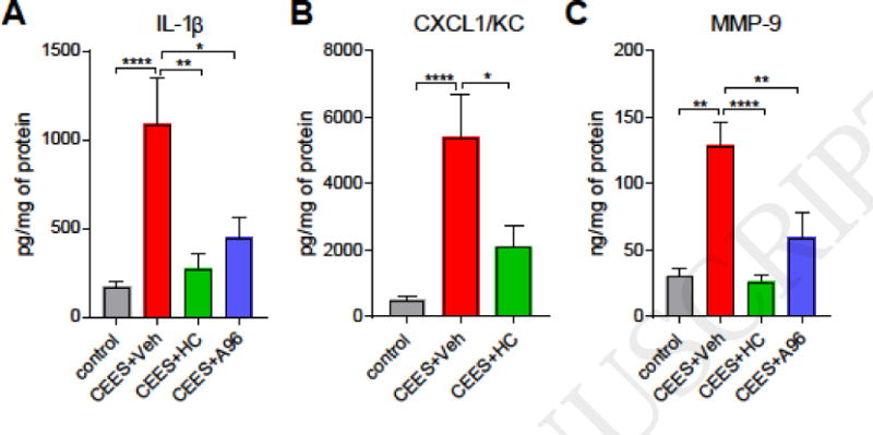
Figure 2. Effects of TRPA1 inhibitor treatment on pro-inflammatory cytokines in ear punch biopsy samples of CEES-exposed mice.

TRPA1 inhibitors (HC-030031, i.p or A-967079, p.o) or vehicle (i.p or p.o) were administered at 1, 8, and 16 h after CEES exposure. (A and B) Quantification of pro-inflammatory cytokines IL-1β and CXCL1/KC in tissue homogenates using ELISA. (C) Levels of MMP-9 in tissue homogenates determined by ELISA. Data are presented as mean ± SEM. n=41/control (dichloromethane only) measurments from all groups; 18/CEES+vehicle (0.5% methyl cellulose), and 9–10/treatment (CEES+HC or CEES+A96). Statistical significance of the difference between the groups was determined by one-way ANOVA followed by Tukey’s multiple comparison post-hoc test. *p < 0.05; **p < 0.01; ****p<0.0001.
