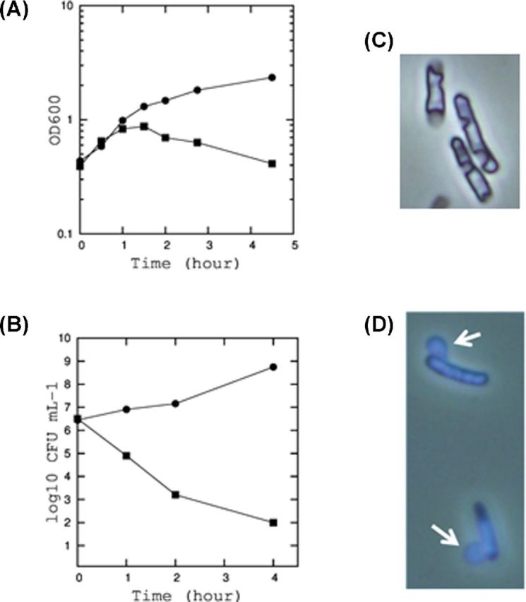
Figure 2.

The growth inhibition and cell lysis by YdfD. (A) Growth curves of E. coli BW25113 expressing YdfD from pBAD24. The cells were incubated in M9 medium. When the optical density at 600 nm reached 0.4, 0.2% arabinose was added (square). Arabinose was also added to the cells containing empty pBAD24 vector as a control (circle). (B) The colony-forming unit (CFU) of cells at 0, 1, 2 and 4 h after the induction of YdfD expression (square) and in control (circle). (C) Cell morphology observed by a phase contrast microscope. (D) Cells stained with DAPI.
