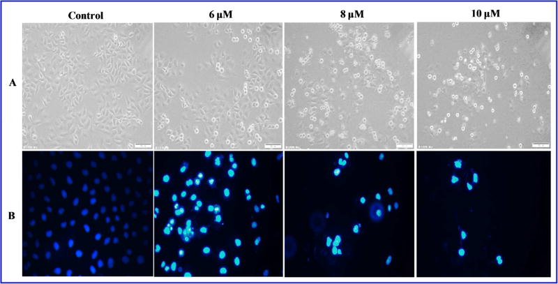
Fig. 6.
(A)The morphological changes of cells were observed under an inverted microscope (magnification, ×400).(B) MGC-803 cells were incubated with indicated concentrations of 1035 for 48 hours and then stained with DAPI. The stained nuclei were then observed under a fluorescentmicroscope (magnification, ×400).

