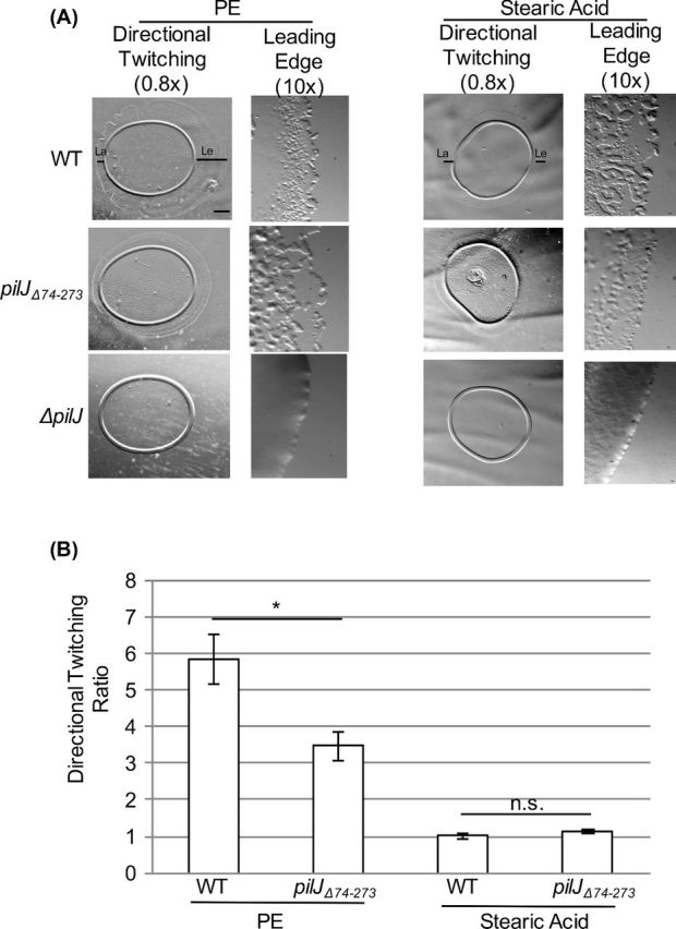
Figure 4.

The putative periplasmic domain of PilJ is not required for directional twitching to PE. (A) Representative images of directional twitching results for the indicated strains. The PE or stearic acid was deposited on the plate to the right of where the P. aeruginosa culture was placed. Black bars on the wild-type strain indicates the leading edge (Le) and lagging edge (La) that were measured to determine the directional twitching ratio. Scale bar = 1mm (0.8× magnification) (B) Directional twitching ratios for both wild type and the pilJΔ74-273 strains. Directional twitching ratios were calculated by dividing the length of the leading edge by the length of the lagging edge. Three independent colonies were analyzed in triplicate. A ratio greater than 2 indicates directional twitching. Significantly different values were determined using a student's t-test (paired, *, P < 0.05).
