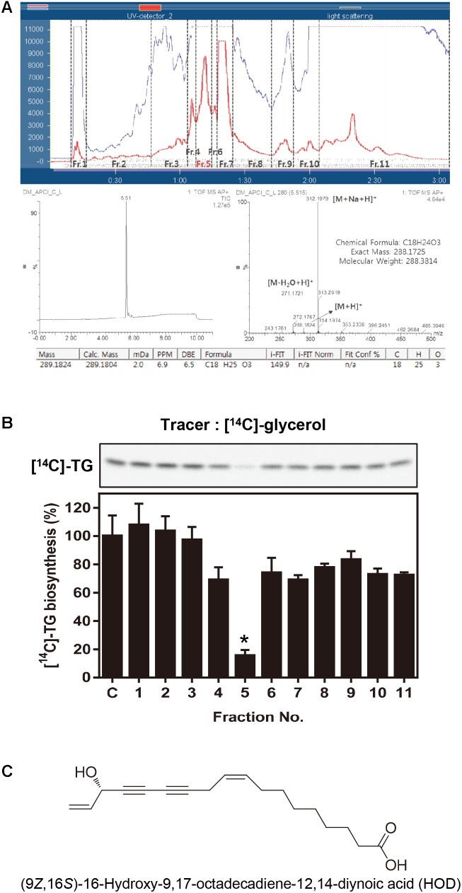
FIGURE 1.
Activity-guided fractionation and identification of HOD. (A) Sepbox separation and UPLC-CAD chromatogram data. (B) 30 μg/ml of eleven fractions were treated in HepG2 cells and evaluated newly biosynthesized TG. Lipid profile analyzed by TLC using [14C]-glycerol as radiolabeled substrates. (C) Chemical structure of HOD. Significance: ∗p < 0.05 vs. control.

