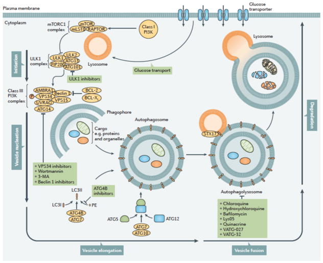
Figure 2. Autophagy can be inhibited at multiple stages.
The process of autophagy is divided into five distinct stages: initiation, vesicle nucleation, vesicle elongation, vesicle fusion and cargo degradation. Nonspecific macroautophagy is initiated by upstream activation through either nutrient starvation or growth factors. Under starvation conditions, a drop in glucose transport results in a release of mTOR inhibition of the ULK1 complex, allowing for the progression of autophagy. The ULK1 complex (comprising ULK1, ULK2, FIP200, ATG101 and ATG13) induces vesicle nucleation which is then mediated by a class III PI3K complex consisting of multiple proteins. Beclin 1, a BCL-2 homology (BH)-3 domain only protein, is phosphorylated by ULK1 and acts as an overall scaffold for the PI3K complex, facilitating localization of autophagic proteins to the phagophore. BCL-2 and BCL-XL interact with Beclin 1 at the BH3 domain to decrease the pro-autophagic activity of Beclin 1 by interrupting the Beclin 1–VPS34 complex formation and decreasing the interaction of Beclin 1 with UVRAG. Additional negative regulation of this process occurs with the phosphorylation of VPS34 (also known as PIK3C3), which decreases its interaction with Beclin 1. In contrast, AMBRA binds Beclin 1 and stabilizes the PI3K complex. ATG14 and UVRAG also bind Beclin 1 to promote interactions between Beclin 1 and VPS34 and phagophore formation. VPS15 is required for optimal VPS34 function by enhancing VPS34 interaction with Beclin 1. The growing double membrane undergoes vesicle elongation to eventually form an autophagasome: a process mediated by two ubiquitin-like conjugation systems. The first system involves the conjugation of phosphatidylethanolamine (PE) to cytoplasmic LC3-I to generate the lipidated form, LC3-II which is facilitated by the protease, ATG4B, and the E1-like enzyme, ATG7, whereby LC3-II is incorporated into the growing membrane. The second conjugation system is mediated again by ATG7 as well as the E2-like enzyme, ATG10, resulting in an ATG5-ATG12 conjugate. Subsequently, the SNARE protein, syntaxin 17 (STX17) facilitates autophagosome fusion with the lysosome, resulting in an autophagolysosome. The low pH of the lysosome results in degradation of the autophagosome contents. This process can be targeted pharmacologically upstream by means of direct ULK1, VPS34, or Beclin 1 inhibition. It can also be targeted by wortmannin and 3-methyladenine (3-MA) which act as PI3K inhibitors. Downstream targets include direct ATG4B inhibitors as well as chloroquine or hydroxychloroquine and bafilomycin, which act to prevent autophagosome fusion with the lysosome. PE, phosphatidylethanolamine.

