Fig. 3.
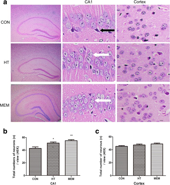
Effect of HT on the histopathological changes in the hippocampus and cortex of APP/PS1 Transgenic Mice. a HE-stained images in the hippocampal CA1 and cortex of APP/PS1, HT and MEM groups. Scale bar = 200 μm. Injured neurons in APP/PS1 mice are darkly stained and exhibited shrunken and triangulated neuronal body (black arrows). Cells are arranged indisorder with a slightly changed cell polarity, neuron loss can also be seen. By contrast, treatment with HT and MEM significantly inhibited the histopathological damage (white arrows). b The hippocampal CA1 in the HT and MEM mice contained significantly more total neurons than did that in the APP/PS1 mice. c The total numbers of neurons in cortex did not significantly differ among the three groups. Data are presented as the mean ± SD. *P < 0.05, **P < 0.01 versus control group. Scale bar = 20 μm, n = 6 per group

