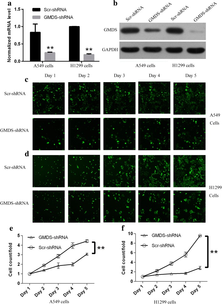
Fig. 2.
Impaired cell proliferation in human lung adenocarcinoma cell lines with GMDS knockdown via Cellomics ArrayScan VTI. a GMDS mRNA level in A549 cells and H1299 cells infected with lentivirus expressing either Scr-shRNA or GMDS-shRNA examined by quantitative real-time PCR (normalized to GAPDH mRNA). Data shown here was one out of three independent experiments (**, p < 0.01). b Relative GMDS protein level in A549 cells and H1299 cells infected with lentivirus expressing either Scr-shRNA or GMDS-shRNA examined by western blot. GAPDH protein was used as internal control. c-d. Representative microscope pictures of A549 cells (c) and H1299 cells (d) infected with lentivirus expressing either Scr-shRNA or GMDS-shRNA at different time points. e-f. Proliferation profiling of A549 cells (e) and H1299 cells (f) infected with lentivirus expressing either Scr-shRNA or GMDS-shRNA for continuous 5 days examined by Cellomics ArrayScan VTI. Histogram shown here was relative fold changes of cell numbers compared to Day 1 and representing the mean ± SEM of three independent experiments (**, p < 0.01)

