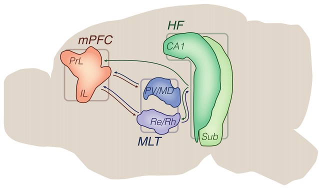
Figure 1.

Principal brain areas and axonal projections of the delimited literature. Mid sagittal view of the rodent brain. Other brain sites are illustrated in subsequent figures (e.g., ventral tegmental area (VTA), basolateral amygdala (BLA), striatum). Arrows from the hippocampal formation (HF) are generically positioned, i.e., no distinction is made between dorsal, intermediate and ventral domains. Drawings are based on the Brain Explorer 3D atlas (Allen Institute). Abbreviations: CA1, cornus ammonis 1; HF, hippocampal formation; IL, infralimbic area; MLT, midline/paramidline thalamus; mPFC, medial prefrontal cortex; PrL, prelimbic area; paraventricular (PV)/mediodorsal thalamus (MD), PV/mediodorsal nuclei; Re/Rh, reuniens/rhomboid nuclei; Sub, subiculum.
