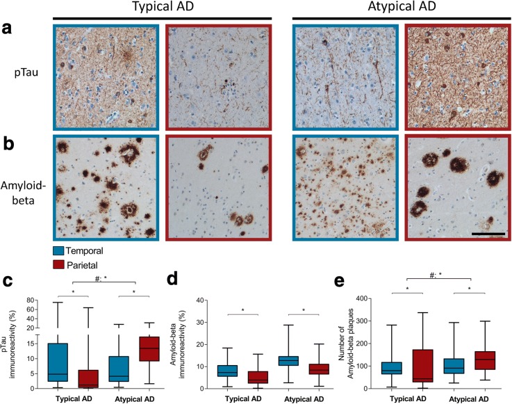
Fig. 2.
pTau and amyloid-beta distribution in typical AD and atypical AD. a In typical AD, the temporal cortex (blue border) shows more immunoreactivity for pTau than the parietal cortex (burgundy border). This distribution is inversed in atypical AD (boxplot in c). b Although both typical and atypical AD show more overall amyloid-beta immunoreactivity in the temporal cortex compared to the parietal region, the atypical AD group shows increased number of amyloid-beta plaques in the parietal compared to temporal section (boxplot in e). Bar represents 100 μm. c, d, and e Boxplots showing pTau immunoreactive area (%), amyloid-beta immunoreactive area (%), and the number of amyloid-beta plaques, respectively, in the temporal and parietal section of both AD phenotypes. Data shown as median (bar), 1st and 3rd quartile (box boundaries), and min to max (error bars). A difference in distribution over the two regions between the two AD phenotypes is indicated by # (Table 5), * p < .0045

