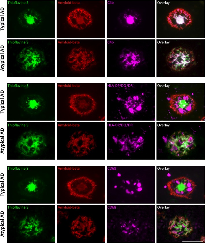
Fig. 7.
Different plaque morphology in the parietal cortex of atypical AD. First row: in typical AD, classical cored plaques in the temporal cortex show an organized staining pattern with a corona showing merely amyloid-beta positivity versus a core positive for thioflavine S, amyloid-beta, and C4b. Second row: in atypical AD, fibrillar plaques in the parietal cortex show co-localization of thioflavine S, amyloid-beta, and C4b in the form of fibrils throughout the whole plaque. Third row: in typical AD, CD68-positive microglia are localized between the core and corona of classical cored plaques. Fourth row: in atypical AD, CD68-positive microglia localization is less organized. Fifth + sixth row: this different distribution within plaques between the two phenotypes also holds for HLA-DP/DQ/DR-positive microglia. Bar is applicable to all images and represents 100 μm

