
Fig. 1.
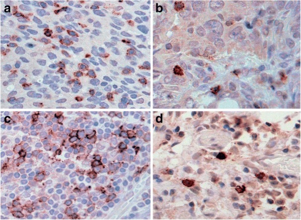
Distinct IHC staining patterns of PD-1 positive cells in the stroma and epithelium of tissue. In ovarian cancer tissue sections, tumor infiltrating lymphocytes (TILS) exhibiting strong membranous and cytoplasmic staining for PD-1 are apparent as clusters (a) or single scattered cells (b). TILS are also seen in aggregates (c) and as single cells (d) in the reactive stroma associated with tumor