
Fig. 3.
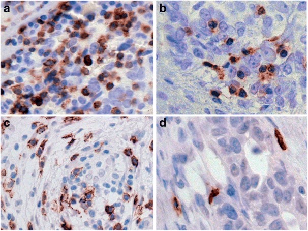
High and low density of TILs in ovarian tumors. IHC staining of T cell subsets in patients’ FFPE tissue sections. CD3 exhibiting diffuse strong staining in clusters of tumor infiltrating lymphocytes (a) versus focal staining in scattered TILs in less dense areas (b). Distribution of CD8 T cells in clusters and as single cells is apparent in c and d respectively