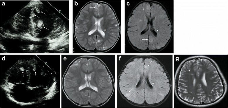
Fig. 1.
Echocardiogram and magnetic resonance imaging. Echocardiogram indicates multiple cardiac rhabdomyomas (arrows) in the ventricles. (a proband; d younger brother). Brain MRI shows multiple cortical tubers (arrows) and small subependymal nodules (*). (b-c proband; e-f younger brother; b, e T2 weighted imaging; c, f T2-tirm-tra-dark-fluid imaging). Axial T2 MRI of the brain demonstrates a central white matter radial migration line (arrow) in the father (g)

